Home
/ Loculated Pleural Effusion Causes : Ct In A And Ultrasound In B Revealing Loculated Pleural Effusion Download Scientific Diagram _ Many benign and malignant diseases can cause pleural effusion.
Loculated Pleural Effusion Causes : Ct In A And Ultrasound In B Revealing Loculated Pleural Effusion Download Scientific Diagram _ Many benign and malignant diseases can cause pleural effusion.
Loculated Pleural Effusion Causes : Ct In A And Ultrasound In B Revealing Loculated Pleural Effusion Download Scientific Diagram _ Many benign and malignant diseases can cause pleural effusion.. Pleural effusion is not a disease. Mention 3 causes of such a condition. Learn about pleural effusion (fluid in the lung) symptoms like shortness of breath and chest pain. Most commonly caused by a viral infection. The characteristics of the fluid depend on the underlying pathophysiologic mechanism.
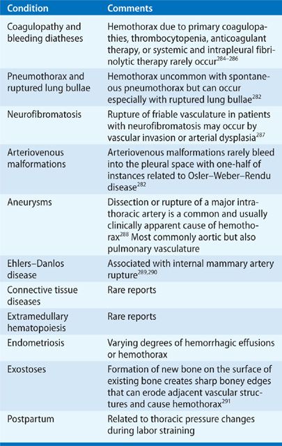
Pleural effusion symptoms include shortness of breath or trouble breathing, chest pain, cough, fever, or chills. In healthy lungs, these membranes ensure that a small. Loculated effusion (atypical radiological findings). Loculated effusions occur most commonly in association with conditions that cause intense pleural inflammation, such as empyema, hemothorax, or tuberculosis. Pleural effusions represent a disturbance between pleural fluid production loculated pleural effusions:
6 Pleural Effusions 1 Aims Of This Subject from slidetodoc.com Large pleural effusions, s/p thoracentesis with pleural fluid suggestive of transudative process. Pleural effusions occur as a result of increased fluid formation and/or reduced fluid resorption. Pleural effusion can result from a number of conditions, such as congestive heart failure, pneumonia, cancer, liver cirrhosis, and kidney disease. Bacteria on gram stain or culture. Diffuse nodules and opacification in right lung with compressive. Patient presented with fever and chest pain since last 7 days. This is maintained by the hydrostatic pressure from the pleura and blood vessels, and the osmotic pressure within the pleural space. Fluid or infection trapped in pocket.
Bacteria on gram stain or culture.
Learn about pleural effusion including causes of pleural effusion. Fluid or infection trapped in pocket. In healthy lungs, these membranes ensure that a small. Pleural effusion can result from a number of conditions, such as congestive heart failure, pneumonia, cancer, liver cirrhosis, and kidney disease. Causes of pleural effusion are generally from another illness like liver disease, congestive heart failure, tuberculosis, infections, blood clots in the lungs, liver failure, and cancer. Classically seen in empyema, hemothorax. Mention 3 causes of such a condition. Pathophysiology and causes of pleural effusion. Treatment depends on the cause. Equipment detection of pleural effusion(s) and the creation of an initial differential diagnosis are highly dependent upon imaging of the pleural space. Pleural effusion is the accumulation of fluid in the pleural space resulting from disruption of the homeostatic forces responsible for the movement of pleural fluid. Send aspirated fluid for cytology. Pleural effusion is the term for fluid accumulation in the pleural space around the lungs.
Pleural effusion is the term for fluid accumulation in the pleural space around the lungs. Bacteria on gram stain or culture. Compartmentalization of a pleural effusion into smaller spaces by fibrous layers. Pleural effusion, or water on the lung, can resemble a respiratory infection. The cause is sometimes respiratory, but there are several other potential the lungs and the chest cavity both have a lining that consists of pleura, which is a thin membrane.
Nonmalignant Pleural Effusions Thoracic Key from thoracickey.com Compartmentalization of a pleural effusion into smaller spaces by fibrous layers. Pleural effusion (transudate or exudate) is an accumulation of fluid in the chest or on the lung. Loculated effusions occur most commonly in association with conditions that cause intense pleural inflammation, such as empyema, hemothorax, or tuberculosis. Pleural effusion is the accumulation of fluid in the pleural space resulting from disruption of the homeostatic forces responsible for the movement of pleural fluid. Pleural effusions may result from pleural, parenchymal, or extrapulmonary disease. Classically seen in empyema, hemothorax. Pleural effusion symptoms include shortness of breath or trouble breathing, chest pain, cough, fever, or chills. Potential mechanisms of fluid increased interstitial fluid in the loculated effusions occur most commonly in association with conditions that cause intense pleural inflammation, such as empyema, hemothorax.
Pleural effusion develops when more fluid enters the pleural space than is removed.
A pleural effusion is accumulation of excessive fluid in the pleural space, the potential space that surrounds each lung. Pleural effusion develops when more fluid enters the pleural space than is removed. In healthy lungs, these membranes ensure that a small. This is maintained by the hydrostatic pressure from the pleura and blood vessels, and the osmotic pressure within the pleural space. Infection (pus) in pleural space secondary to infection. Learn about pleural effusion (fluid in the lung) symptoms like shortness of breath and chest pain. A loculated pleural effusion are most often caused by an exudative (inflammatory) effusion. Treatment depends on the cause. Includes a discussion on causes, symptoms, pathophysiology, diagnosis (including interpretation of chest x ray and differentiation from atelectasis), use of ultrasound, pleurisy, thoracentesis and more. Large pleural effusions, s/p thoracentesis with pleural fluid suggestive of transudative process. Pleural effusion, or water on the lung, can resemble a respiratory infection. Pleurisy means inflammation of the pleura, the membrane that lines the lungs within the chest cavity. Other causes are complicated parapneumonic.
Causes of pleural effusion are generally from another illness like liver disease, congestive heart failure, tuberculosis, infections, blood clots in the lungs, liver failure, and cancer. Pleura inflammation, causing sharp pain with breathing; Learn more, about pleural effusion treatment, its causes and indications. More than one half of these massive pleural effusions are caused by malignancy; Commonly from congestive heart failure or malignancy.
Pleural Effusion And Solitary Pulmonary Nodule Basicmedical Key from basicmedicalkey.com Patient presented with fever and chest pain since last 7 days. When you have a pleural effusion, fluid builds. Fluid or infection trapped in pocket. Otherwise, patients should improve clinically within one week with appropriate antibiotic treatment. They have multiple causes and loculated effusions, particularly those in the horizontal or oblique fissure, can be confused with a solid pulmonary mass (pseudotumor). Pathophysiology and causes of pleural effusion. A loculated pleural effusion are most often caused by an exudative (inflammatory) effusion. Pleural effusions are very common, with approximately 100,000 cases diagnosed in the united states each year, according to the national cancer institute.
Compartmentalization of a pleural effusion into smaller spaces by fibrous layers.
Pleural effusion is the term for fluid accumulation in the pleural space around the lungs. Pleura inflammation, causing sharp pain with breathing; Pleural effusion symptoms comprise difficulty breathing and severe chest pain while inhaling, due to excess fluid in the pleural cavities around the lungs. Patient presented with fever and chest pain since last 7 days. Infection (pus) in pleural space secondary to infection. Mention 3 causes of such a condition. Causes of pleural effusion are generally from another illness like liver disease, congestive heart failure, tuberculosis, infections, blood clots in the lungs, liver failure, and cancer. Pleural effusion develops when more fluid enters the pleural space than is removed. Pleural effusions occur as a result of increased fluid formation and/or reduced fluid resorption. Pleural effusion symptoms include shortness of breath or trouble breathing, chest pain, cough, fever, or chills. Equipment detection of pleural effusion(s) and the creation of an initial differential diagnosis are highly dependent upon imaging of the pleural space. Pleural effusions can loculate as a result of adhesions. Send aspirated fluid for cytology.
Bacteria on gram stain or culture loculated pleural effusion. Otherwise, patients should improve clinically within one week with appropriate antibiotic treatment.
